عضویت
عضویت  ورود اعضا
 راهنمای خرید


QImaging Retiga 60000 pages

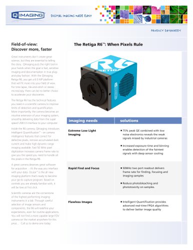
PRODUCT DATASHEET
Large Field of View, High Resolution
Fluorescence Snapshot Camera
Fluorescence Imaging
with a Larger Perspective
The vast majority of fluorescence cameras
used today capture only 24% of the usable
Field of View (FOV) on the microscope.
This small FOV significantly reduces the
amount of data collected by a single frame,
limiting the number of cells monitored
simultaneously, increasing the number of
frames required for whole slide imaging and
overall slowing down the throughput of a
lab. To counter this FOV challenge, many
scientists resort to de-magnifying optical
adapters or lower magnifications
but at the cost of significantly reduced
image resolution.
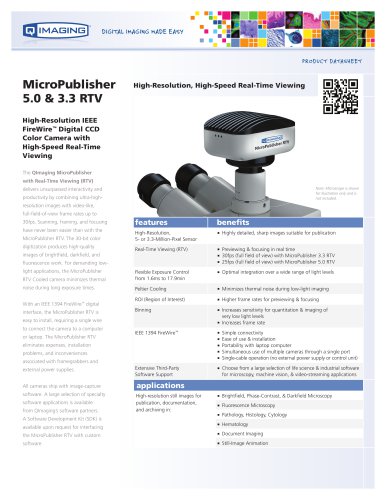
With the Retiga 6000 from QImaging,
capturing the largest FOV is possible without
compromising on resolution or sensitivity.
With 6.05 million pixels and a 16mm sensor
diagonal, the Retiga 6000 exploits the full
microscope FOV, capturing twice the image
area than most standard fluorescence
cameras. Additionally, the small 4.54μm
pixels maintain Nyquist sampling with lower
magnifications and new high NA objectives,
allowing the camera to benefit from the
boost in optical resolution and image area.
In the past, smaller pixels resulted in
substantially less sensitivity, consequently
limiting their use in fluorescence microscopy.
However, with low noise camera electronics,
high quantum efficiency of 75% and
low dark current, the Retiga 6000 offers
superior resolution without compromising
on sensitivity.
features
See More of What You
Are Missing
t 16mm sensor diagonal
<
Switch Between Low and
High Magnifications without
Compromise
t 6.05 Mega Pixels with 4.54μm
t pixel pitch
<
Less Light? Not a Problem
t 75% QE combined with
t low noise electronics
<
Easy Compatibility with
Virtually Any Windows PC
t USB 2.0
t QCapture Pro™
<
<
An unequivocal advance in fluorescence
documentation, the Retiga 6000 allows
users to see larger areas in greater detail and
with higher clarity than before, helping to
push forward throughput and productivity
in the lab.
benefits
tCapture twice the image field of standard
microscopy CCD cameras*
<tSimultaneously monitor twice as many cells for
rare event detection, phenotyping, imaging
cytometry, and cell cycle documentation
<tReduce the number of frames by half when
scanning whole slides
<
tLarge numbers of small pixels are ideal for low
magnification, large FOV work
<tSmall pixels take advantage of the increased
optical resolution with new high NA objectives
<tUse binning to combine pixels and improve
sensitivity at high magnifications
<
tReduce exposure times and the negative effects
of bleaching and phototoxicity
<tAchieve higher resolution imaging without
compromising on sensitivity
<tImage low luminescence signals over long
periods
<
tNo cards to install, plug and play simplicity**
tIncluded image acquisition software QCapture
Pro, combines a simple work-flow with basic
analysis tools
<
<
*Based on comparison with other microscopy cameras utilizing a 2/3” sensor.
**Minimum computer specifications required to ensure performance. Please see the QImaging website for more details.